
In a further two cases, where no neck nodes were seen, a histological diagnosis of sarcoidosis was made from biopsy of diffusely abnormal parotid gland tissue.Ĭonclusions: Given the advantages of cervical diagnosis in terms of invasiveness and economy compared to mediastinal alternatives, it is suggested that where the expertise for core biopsy of normal sized cervical lymph nodes is readily available, the technique may be considered as a first line investigation for the diagnosis of sarcoidosis. Lymph nodes are part of the lymphatic system, which is a complex network of nodes and vessels. Lymph nodes are small, round or bean-shaped glands that act like filters. Nevertheless histological examination revealed non-caseating granulomas in all cases.
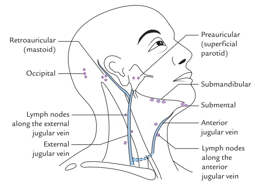
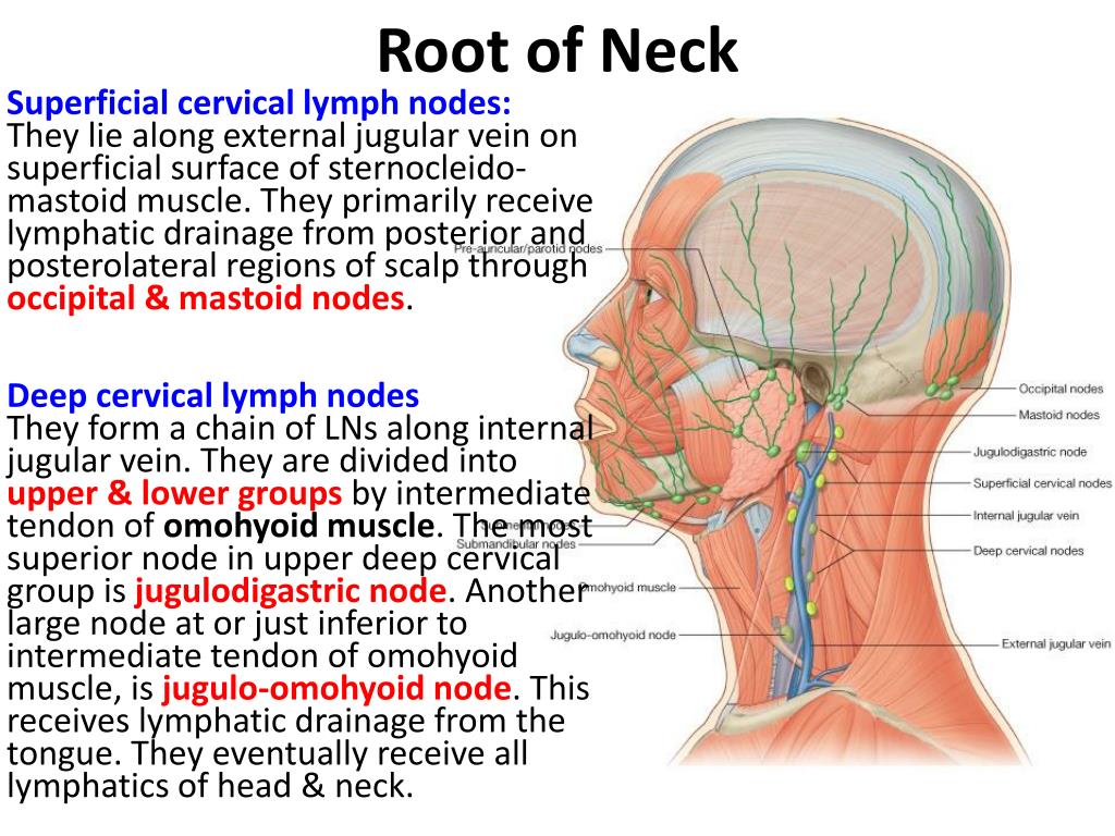
#CERVICAL LYMPH NODES SERIES#
It is emphasised that the cervical lymph nodes in this series were not particularly enlarged, short axis dimensions being under 10mm in 6 of the 7 cases biopsied, and that these sub-centimeter lymph nodes did not have any specific sonographic appearances to mark them as pathological. Results: A diagnosis of sarcoidosis was made in all cases where a core biopsy was attempted. The term lymphadenopathy strictly speaking refers to. Where no cervical node suitable for biopsy was seen, the parotid glands were evaluated and biopsied if abnormal. Cervical lymphadenopathy refers to lymphadenopathy of the cervical lymph nodes (the glands in the neck). Typically these patients had no clinically apparent neck nodes. The source of the infection may not be in the neck itself but may be from the areas of. Cervical lymph nodes are the most common location for residual and recurrent PTC. Methods: Nine patients were referred for sonographic evaluation of the head and neck to avoid the use of more invasive and expensive tests such as endobronchial ultrasound and mediastinoscopy. Lymph nodes in the neck may also be enlarged from bacterial infections. We describe 9 cases where histological diagnosis was made with US guided core biopsy to confirm clinically and radiologically suspected sarcoidosis.

The diagnostic value of neck ultrasound with core biopsy for histological confirmation as a less invasive approach was evaluated. Here we show that a brain-to-cervical lymph node (CLN) pathway is involved. Aims and Objectives: Sarcoidosis is a multi-system granulomatous disease of uncertain aetiology. Abstract After stroke, peripheral immune cells are activated and these systemic responses may amplify brain damage, but how the injured brain sends out signals to trigger systemic inflammation remains unclear.


 0 kommentar(er)
